
What are Glial Tumors (Gliomas)?
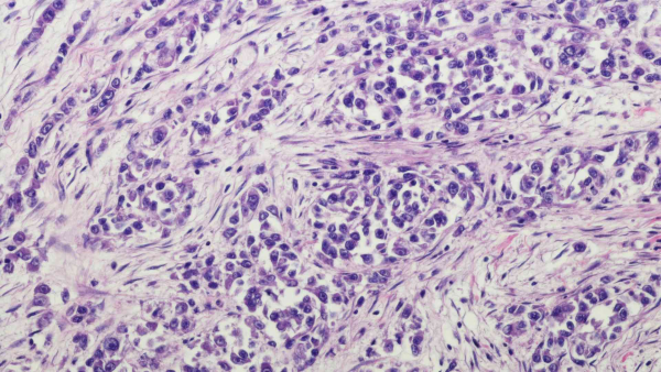
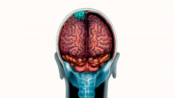
Glial tumors are tumors that originate from the glial cells, which form the supporting tissue of the brain or spinal cord. Although it varies by age, approximately 80% of malignant brain tumors are glial tumors. The brain and spinal cord are composed of “nerve cells (neurons)” and the supporting tissue called “glial cells.” Glial cells support, protect, and nourish neurons.
Glial tumors usually arise as a result of abnormal cell growth originating from glial cells. These tumors can be either benign (non-cancerous) or malignant (cancerous). Benign glial tumors generally have a better prognosis and are often treatable with surgical intervention. However, malignant glial tumors exhibit more aggressive growth and have the potential to spread cancerous cells to surrounding tissues.
What is a Genetic Test for Glial Tumors (Gliomas)?
Genetic testing for glial tumors includes a series of laboratory tests used to examine the molecular structure and genetic characteristics of a patient’s tumor. These tests are important for better understanding the tumor type, its spread, response to treatment, and prognosis. “Genetic tests for glial tumors” may include the following:
-
Genetic Profile Analysis: DNA is extracted from a biopsy sample taken from the tumor tissue or from the entire excised tumor tissue. Using next-generation sequencing (NGS), genetic mutations that may guide diagnosis, prognosis, and treatment decisions are analyzed.
-
Immunohistochemistry (IHC) Analysis: This technique is used to determine the presence and quantity of specific proteins and molecules in the tumor. It can help identify the type and aggressiveness of the tumor.
-
FISH (Fluorescence In Situ Hybridization): This method is used to detect specific genetic changes. Appropriate thickness sections are taken from the tissue, and the cancerous region is identified in the pathology lab. FISH testing is then performed on genes associated with gliomas in the cancerous area.
-
Tumor Mutation Panel: These are genetic panel tests performed using next-generation sequencing (NGS) technology to simultaneously test for various genetic changes. The panel may be performed using tumor tissue or through liquid biopsy by extracting serum from a blood sample.
This test evaluates whether mutations exist in numerous genes, while also simultaneously calculating microsatellite instability and tumor mutation burden.
These panels provide a detailed analysis of the tumor’s genetic profile. They help identify the unique characteristics of the tumor in the patient, which is crucial for determining “personalized treatment” options and prognosis. Different centers may offer tests that cover varying numbers of genes.
In our center, we use a panel that includes a broad range of genetic mutations and also evaluates Tumor Mutation Burden (TMB), PDL/PDL1, and Microsatellite Instability.
This panel also simultaneously analyzes polymorphisms related to the toxic effects and efficacy of classical chemotherapeutic agents in patients. For patients diagnosed with glioma cancer, it is recommended to consult an oncologist and a genetics specialist to evaluate treatment options.
These genetic tests can help determine the tumor’s sensitivity to treatment options and the patient’s prognosis. Certain genetic mutations, in particular, may make tumors more responsive to specific drugs or targeted therapies. Therefore, genetic testing for glial tumors is important for personalizing patients’ treatment plans.
Some Genetic Tests Used for Glial Tumors (Gliomas) and Brain Tumors
-
IDH1 and IDH2 (Codons 132 and 172)
-
ATRX (Sequencing analysis)
-
P53 (TP53) (Sequencing analysis)
-
EGFR (Exons 18, 19, 20, 21)
-
CDKN2A (Sequencing analysis)
-
BRAF Mutations (Sequencing analysis)
-
1p19q (FISH)
-
PDL1 Expression (Immunohistochemistry)
-
PIK3CA (Next-Generation Sequencing - NGS) (Exon 9: E542 and E545; Exon 20: H1047)
-
MTOR (for inhibitors)
Types and Characteristics of Glial Tumors (Gliomas)
Tumors resulting from the abnormal growth or proliferation of glial cells are generally classified as glial tumors and may include:
-
Astrocytomas: Astrocytomas originate from glial cells called astrocytes. They can occur in various parts of the brain. Astrocytomas can sometimes be benign (non-cancerous), but some may be malignant (cancerous) and grow rapidly. The prognosis varies depending on the tumor grade.
-
Oligodendrogliomas: This type of glial tumor originates from glial cells called oligodendrocytes. They are usually found in the brain and are often benign.
-
Ependymomas: Ependymomas are glial tumors that originate from ependymal cells in the brain and spinal cord. They are typically located in the spinal canal and brain ventricles. There are both slow-growing and fast-growing types.
These glial tumors often lead to neurological symptoms. Their clinical course and treatment approaches vary depending on the type, size, and location of the tumor, the patient’s age, and overall health condition. Treatment options may include surgery, radiotherapy, chemotherapy, immunotherapy, and targeted therapies.
What Are the Symptoms of Glial Tumors (Gliomas)?
Since glial tumors originate from the glial cells of the brain or spinal cord, the symptoms generally manifest as issues related to the functions of these organs. Symptoms may vary depending on the tumor’s type, size, location, and the patient’s general health status. Common symptoms of glial tumors include:
-
Headache: Glial tumors are often associated with headaches. These headaches are typically more pronounced in the mornings and tend to be persistent.
-
Neurological Symptoms: Depending on the tumor’s location, patients may experience various neurological symptoms. These may include loss of balance, coordination difficulties, memory problems, forgetfulness, muscle weakness, speech disorders, paralysis, numbness, or tingling sensations.
-
Visual Problems: The tumor may press on the optic nerves or visual pathways, leading to visual disturbances. These may include double vision, vision loss, or changes in the visual field.
-
Seizures: Glial tumors may sometimes cause seizures. The severity and frequency of seizures depend on the tumor’s characteristics.
-
Altered Consciousness: Particularly in cases of large tumors or tumors causing edema, altered consciousness may range from mild confusion to loss of consciousness.
-
Nausea and Vomiting: Brain tumors may increase intracranial pressure, leading to nausea and vomiting.
-
Personality and Behavioral Changes: Depending on the affected areas, personality changes, irritability, or emotional instability may occur.
It is important to note that these symptoms may also be associated with many other conditions. Therefore, if an individual experiences such symptoms, it is essential to consult a healthcare professional. As with all cancers, early diagnosis and treatment of “glial tumors” will significantly impact the disease’s course.